Belle Rodrequez
  • Blog
  • About
  • Contact

Do Fallen Arches Have To Have Surgery?

7/2/2017

 
Overview

Flat Feet

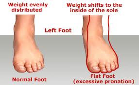
Fallen arches, also know as having flat feet, or pes planus, refers to the collapsing of the foot?s arch so that it is no longer curved properly or adequately supportive. The condition can cause foot pain, fatigue and affect gait, which can create strain on the knees, ankles legs and hips. Fallen arches can occur naturally from birth, or can be attributed to the rolling in of the foot and ankle while running (overpronation).

Causes

The arches of most individuals are fully developed by the age of 12 to 13. While some people are born with flat arches, for others, the arches fall over time. The tibial tendon, which runs along the inside of the ankle from above the ankle to the arch, can weaken with age and with heavy activity. The posterior tendon, main support structure for the arch, can become inflamed (tendonitis) or even tear if overloaded. For women, wearing high heels can affect the Achilles tendon and alter the structure and function of the ankle. The posterior tibial tendon may compensate for this stress and break down, causing the arches to fall. Obesity is another contributing factor, as well as a serious injury to the ankle or foot, arthritis and bad circulation such as occurs with diabetes.

Symptoms

Not everyone who has flat feet experiences symptoms. Others, however, feel persistent pain in their feet and wearing shoes can prove additionally painful. Others only feel symptoms when they walk more than normal, go jogging or participate in a sport that involves running or kicking.

Diagnosis

You can test yourself to see if you have flat feet or fallen arches by using a simple home experiment. First, dip your feet in water. Then step on a hard flat surface, like a dry floor or a piece of paper on the floor, where your footprints will show. Step away and examine your foot prints. If you see complete/full imprints of your feet on the floor, you may have fallen arches. However, it?s important to seek a second option from a podiatrist if you suspect you have fallen arches so they can properly diagnose and treat you.

fallen arches surgery

Non Surgical Treatment

During walking and running, there is a small natural inward drop (slight pronation) that is part of the spring and propulsion. Allowing exaggerated sagging is like rounding your shoulders too much. Legs and feet have posture that you can control yourself. Use your own muscles and get free built-in exercise and arch support all day, and stop painful poor positioning. Some people with existing abnormality or growths in the ball of the foot will roll inward (or outward) to get the pressure off the deformed area because standing straight hurts. See your doctor first. Remember, don't force. If it hurts, it's wrong. All you are doing is learning how to stand neutral, not tilted so much that you compress the joints. The concept is to hold your feet in the same healthful position that shoe supports would. It is like an ice skater holds their skates straight at the ankle, not angled.

Surgical Treatment

Acquired Flat Foot

Surgery is typically offered as a last resort in people with significant pain that is resistant to other therapies. The treatment of a rigid flatfoot depends on its cause. Congenital vertical talus. Your doctor may suggest a trial of serial casting. The foot is placed in a cast and the cast is changed frequently to reposition the foot gradually. However, this generally has a low success rate. Most people ultimately need surgery to correct the problem. Tarsal coalition. Treatment depends on your age, extent of bone fusion and severity of symptoms. For milder cases, your doctor may recommend nonsurgical treatment with shoe inserts, wrapping of the foot with supportive straps or temporarily immobilizing the foot in a cast. For more severe cases, surgery is necessary to relieve pain and improve the flexibility of the foot. Lateral subtalar dislocation. The goal is to move the dislocated bone back into place as soon as possible. If there is no open wound, the doctor may push the bone back into proper alignment without making an incision. Anesthesia is usually given before this treatment. Once this is accomplished, a short leg cast must be worn for about four weeks to help stabilize the joint permanently. About 15% to 20% of people with lateral subtalar dislocation must be treated with surgery to reposition the dislocated bone.

Heel Painfulness The Major Causes, Signals And Therapy Choices

6/29/2017

 
Overview

Painful Heel

The heel bone is the largest of the 26 bones in the human foot, which also has 33 joints and a network of more than 100 tendons, muscles, and ligaments. Like all bones, it is subject to outside influences that can affect its integrity and its ability to keep us on our feet. Heel pain, sometimes disabling, can occur in the front, back, or bottom of the heel.

Causes

There are two different categories of heel pain. The first is caused by over-use repetitive stress which refers to a soreness resulting from too much impact on a specific area of the foot. This condition, often referred to as "heel pain syndrome," can be caused by shoes with heels that are too low, a thinned out fat pad in the heel area, or from a sudden increase in activity. Plantar fasciitis, a very common diagnosis of heel pain, is usually caused from a biomechancial problem, such as over-pronation (flat feet). The plantar fascia is a broad band of fibrous tissue that runs along the bottom surface of the foot, from the heel through the midfoot and into the forefoot. Over-pronation can cause the plantar fascia to be excessively stretched and inflamed, resulting in pain in the heel and arch areas of the foot. Often the pain will be most intense first thing in the morning or after a prolonged period of rest. The pain will gradually subside as the day progresses.

Symptoms

Sever?s Disease. This is a condition that occurs in 10 - 15 year old children, predominantly boys and is associated with running and repetitive jumping. It is also associated with flimsy footwear that kids may wear. It occurs when the Achilles tendon continually pulls on the apophysis of the calcaneum and does not allow for it to fuse with the body of the calcaneum. Calcaneal enthesopathy. This occurs when there is repetitive trauma at the attachment of the Achilles tendon, resulting in a spur from the calcaneum up into the Achilles tendon. It is usually visualized on x-ray and may be tender if there is an associated bursitis or tendonitis. "Pump Bump". Also known as Haglund?s Deformity, this is a bony enlargement that exists on the back of the heel - usually related to a congenital abnormality or with chronic bursitis, causing a thickening. There may have already been trauma or pressure from footwear. Treatment is usually protection of the bump and correct footwear. Associated with a symmetrical swelling at the base of the Achilles tendon. It is usually related to repetitive trauma or inappropriate footwear. It is often red and hot in the early stages. Treatment is usually to correct the footwear, provide padding and treat the local symptoms e.g. ice, rest, physiotherapy and cortisone injection. Fat Pad Syndrome. Direct contact with the base of the heel may result in trauma to the fat pad. Related to obesity, training on hard surfaces, uneven grounds, poor shoes especially overlarge shoes which can cause shearing forces on the heel. These conditions are renowned for taking a long time to recover - usually many months.

Diagnosis

In most cases, your GP or a podiatrist (a specialist in foot problems and foot care) should be able to diagnose the cause of your heel pain by asking about your symptoms and medical history, examining your heel and foot.

Non Surgical Treatment

Calf stretch, silicone Heel cups, ice, night splint, physical therapy. Sometimes custom orthotics are beneficial in long standing cases. Steroid injections have been used and although they temporarily relieve the pain, the pain usually returns within a short period of time. Plantar fasciitis tends to go away in 90% of all people in time. It can take 12-18 months for all the pain to resolve. If the pain continues after adequate treatment, high frequency shock wave therapy (OssaTron) has been found to be beneficial, unfortunately most insurance companies do not cover this procedure.

Surgical Treatment

Extracorporeal shockwave therapy (EST) is a fairly new type of non-invasive treatment. Non-invasive means it does not involve making cuts into your body. EST involves using a device to deliver high-energy soundwaves into your heel. The soundwaves can sometimes cause pain, so a local anaesthetic may be used to numb your heel. It is claimed that EST works in two ways. It is thought to have a "numbing" effect on the nerves that transmit pain signals to your brain, help stimulate and speed up the healing process. However, these claims have not yet been definitively proven. The National Institute for Health and Care Excellence (NICE) has issued guidance about the use of EST for treating plantar fasciitis. NICE states there are no concerns over the safety of EST, but there are uncertainties about how effective the procedure is for treating heel pain. Some studies have reported that EST is more effective than surgery and other non-surgical treatments, while other studies found the procedure to be no better than a placebo (sham treatment).

ankle strap for heels

Prevention

Painful Heel

Make sure you wear appropriate supportive shoes. Don't over-train in sports. Make sure you warm up, cool down and undertake an exercise regime that helps maintain flexibility. Manage your weight, obesity is a factor in causing plantar fasciitis. Avoid walking and running on hard surfaces if you are prone to pain. You should follow the recognized management protocol "RICED" rest, ice, compression, elevation and diagnosis. Rest, keep off the injured ankle as much as possible. Ice, applied for 20 minutes at a time every hour as long as swelling persists. Compression, support the ankle and foot with a firmly (not tightly) wrapped elastic bandage. Elevation, keep foot above heart level to minimize bruising and swelling. Diagnosis. Consult a medical professional (such as a Podiatrist or doctor) especially if you are worried about the injury, or if the pain or swelling gets worse. If the pain or swelling has not gone down significantly within 48 hours, also seek treatment. An accurate diagnosis is essential for proper rehabilitation of moderate to severe injuries.

Genetic Leg Length Discrepancy

6/28/2017

 
Overview

Surgical lengthening of the shorter extremity (upper or lower) is another treatment option. The bone is lengthened by surgically applying an external fixator to the extremity in the operating room. The external fixator, a scaffold-like frame, is connected to the bone with wires, pins or both. A small crack is made in the bone and tension is created by the frame when it is "distracted" by the patient or family member who turns an affixed dial several times daily. The lengthening process begins approximately five to ten days after surgery. The bone may lengthen one millimeter per day, or approximately one inch per month. Lengthening may be slower in adults overall and in a bone that has been previously injured or undergone prior surgery. Bones in patients with potential blood vessel abnormalities (i.e., cigarette smokers) may also lengthen more slowly. The external fixator is worn until the bone is strong enough to support the patient safely, approximately three months per inch of lengthening. This may vary, however, due to factors such as age, health, smoking, participation in rehabilitation, etc. Risks of this procedure include infection at the site of wires and pins, stiffness of the adjacent joints and slight over or under correction of the bone?s length. Lengthening requires regular follow up visits to the physician?s office, meticulous hygiene of the pins and wires, diligent adjustment of the frame several times daily and rehabilitation as prescribed by your physician.Leg Length Discrepancy

Causes

Limb-length conditions can result from congenital disorders of the bones, muscles or joints, disuse or overuse of the bones, muscles or joints caused by illness or disease, diseases, such as bone cancer, Issues of the spine, shoulder or hip, traumatic injuries, such as severe fractures that damage growth plates.

Symptoms

Often there are few or no symptoms prior to the age of 25-35. The most common symptom is chronic lower back pain, but also is frequently middle and upper back pain. Same-sided and repeated injury or pain to the hip, knee and/or ankle is also a hallmark of a long-standing untreated LLD. It is not uncommon to have buttock or radiating hip pain that is non-dermatomal (not from a disc) and tends to go away when lying down.

Diagnosis

A doctor will generally take a detailed medical history of both the patient and family, including asking about recent injuries or illnesses. He or she will carefully examine the patient, observing how he or she moves and stands. If necessary, an orthopedic surgeon will order X-ray, bone age determinations and computed tomography (CT) scans or magnetic resonance imaging (MRI).

Non Surgical Treatment

Treatment depends on the amount and cause of the leg length discrepancy as well as the age of your child. Typically, if the difference is less than 2 cm we don?t recommend immediate treatment. We may recommend that your child wear a heel lift in one shoe to make walking and running more comfortable. If the leg length discrepancy is more significant, your doctor may recommend surgery to shorten or lengthen a leg. The procedure used most often to shorten a leg is called epiphysiodesis.

LLD Shoe Inserts

what shoes make you taller

Surgical Treatment

Surgical lengthening of the shorter extremity (upper or lower) is another treatment option. The bone is lengthened by surgically applying an external fixator to the extremity in the operating room. The external fixator, a scaffold-like frame, is connected to the bone with wires, pins or both. A small crack is made in the bone and tension is created by the frame when it is "distracted" by the patient or family member who turns an affixed dial several times daily. The lengthening process begins approximately five to ten days after surgery. The bone may lengthen one millimeter per day, or approximately one inch per month. Lengthening may be slower in adults overall and in a bone that has been previously injured or undergone prior surgery. Bones in patients with potential blood vessel abnormalities (i.e., cigarette smokers) may also lengthen more slowly. The external fixator is worn until the bone is strong enough to support the patient safely, approximately three months per inch of lengthening. This may vary, however, due to factors such as age, health, smoking, participation in rehabilitation, etc. Risks of this procedure include infection at the site of wires and pins, stiffness of the adjacent joints and slight over or under correction of the bone?s length. Lengthening requires regular follow up visits to the physician?s office, meticulous hygiene of the pins and wires, diligent adjustment of the frame several times daily and rehabilitation as prescribed by your physician.

Addressing Mortons Neuroma

6/5/2017

 
Overview

Morton neuromaMorton's neuroma, also called Morton's metatarsalgia, Morton's disease, Morton's neuralgia, Morton metatarsalgia, Morton nerve entrapment, plantar neuroma, or intermetatarsal neuroma is a benign (non-cancerous) growth of nerve tissue (neuroma) that develops in the foot, usually between the third and fourth toes (an intermetatarsal plantar nerve, most commonly of the third and fourth intermetatarsal spaces). It is a common, painful condition.

Causes

There are orthoses and corrective shoes that can effectively alleviate disturbances to foot mechanics. A podiatric physician can prescribe the best corrective footwear and shoe inserts for all activities, work, exercise, play, walking, shopping and more, based on an analysis of the patient?s foot and his or her lifestyle. Improper footwear. Podiatric physicians have long believed that constricting, narrow, poor-fitting shoes with a tight or pointed toe box tend to compress the end of the foot, leading to abnormal motion of the foot and to excessive pressure in the area of the nerve. High-heeled shoes are a particular culprit here, since they put pressure on the area around wearer?s toes and the ball of the foot.

Symptoms

People with Morton's neuroma usually complain of pain that can start in the ball of the foot and shoot into the affected toes. However, some people just have toe pain. There may also be burning and tingling of the toes. The symptoms are usually felt up the sides of the space between two toes. For example, if the nerve between the third and fourth long bones (metatarsals) of the right foot is affected, the symptoms will usually be felt up the right-hand side of the fourth toe and up the left-hand side of the third toe. Some people describe the pain that they feel as being like walking on a stone or a marble. Symptoms can be made worse if you wear high-heeled shoes. The pain is relieved by taking your shoe off, resting your foot and massaging the area. You may also experience some numbness between the affected toes. Your affected toes may also appear to be spread apart, which doctors refer to as the 'V sign'. The symptoms can vary and may come and go over a number of years. For example, some people may experience two attacks of pain in a week and then nothing for a year. Others may have regular and persistent (chronic) pain.

Diagnosis

Patients with classic Morton?s neuroma symptoms will have pain with pressure at the base of the involved toes (either between the 2nd and 3rd toes, or between the 3rd and 4th toes). In addition, squeezing the front of the foot together can exacerbate symptoms. As well, they may have numbness on the sides of one toe and the adjacent toe, as this corresponds with the distribution of the involved nerve.

Non Surgical Treatment

Common treatments involve wearing different shoes or using arch supports. Resting the foot, massaging the toes and using an ice pack may work for some people. A GP or a podiatrist (foot specialist) may also recommend anti-inflammatory painkillers or a course of steroid injections. Numbing injections, in which alcohol and a local anaesthetic are injected into the affected area of the foot, may also be effective. In extreme cases, when the condition does not respond to treatment, day case surgery may be needed.Morton

Surgical Treatment

About one person in four will not require any surgery for Morton's neuroma and their symptoms can be controlled with footwear modification and steroid/local anaesthetic injections. Of those who choose to have surgery, about three out of four will have good results with relief of their symptoms. Recurrent or persisting (chronic) symptoms can occur after surgery. Sometimes, decompression of the nerve may have been incomplete or the nerve may just remain 'irritable'. In those who have had cutting out (resection) of the nerve (neurectomy), a recurrent or 'stump' neuroma may develop in any nerve tissue that was left behind. This can sometimes be more painful than the original condition.

Prevention

It is not always possible to prevent a Morton's neuroma. However, you probably can reduce your risk by wearing comfortable shoes that have low heels, plenty of toe space and good arch support.

Shoe Lifts The Best Solution To Leg Length Imbalances

2/27/2016

 
There are not one but two different types of leg length discrepancies, congenital and acquired. Congenital implies that you are born with it. One leg is anatomically shorter compared to the other. As a result of developmental phases of aging, the brain picks up on the gait pattern and identifies some variation. Your body usually adapts by tilting one shoulder to the "short" side. A difference of under a quarter inch is not very abnormal, does not need Shoe Lifts to compensate and in most cases doesn't have a profound effect over a lifetime.

Leg Length Discrepancy Shoe Lift

Leg length inequality goes largely undiscovered on a daily basis, however this problem is very easily remedied, and can reduce a number of instances of lumbar pain.

Treatment for leg length inequality commonly involves Shoe Lifts. Many are economical, ordinarily being less than twenty dollars, in comparison to a custom orthotic of $200 or higher. When the amount of leg length inequality begins to exceed half an inch, a whole sole lift is generally the better choice than a heel lift. This prevents the foot from being unnecessarily stressed in an abnormal position.

Lumbar pain is easily the most prevalent ailment impacting men and women today. Over 80 million men and women have problems with back pain at some stage in their life. It is a problem that costs companies vast amounts of money annually due to lost time and production. Innovative and superior treatment solutions are constantly sought after in the hope of reducing the economical influence this issue causes.

Shoe Lift

People from all corners of the world suffer the pain of foot ache due to leg length discrepancy. In a lot of these situations Shoe Lifts are usually of very useful. The lifts are capable of eliminating any discomfort in the feet. Shoe Lifts are recommended by countless professional orthopaedic practitioners".

So that you can support the body in a well-balanced manner, your feet have a significant task to play. In spite of that, it is often the most neglected area in the human body. Some people have flat-feet meaning there is unequal force exerted on the feet. This causes other body parts such as knees, ankles and backs to be affected too. Shoe Lifts make sure that correct posture and balance are restored.

Are Shoe Lifts The Answer To Leg Length Discrepancy

2/24/2016

 
There are not one but two unique variations of leg length discrepancies, congenital and acquired. Congenital indicates that you are born with it. One leg is anatomically shorter compared to the other. Through developmental periods of aging, the human brain senses the stride pattern and identifies some variation. The body typically adapts by dipping one shoulder over to the "short" side. A difference of under a quarter inch isn't grossly uncommon, doesn't need Shoe Lifts to compensate and typically won't have a profound effect over a lifetime.

Leg Length Discrepancy Shoe Lift

Leg length inequality goes mainly undiagnosed on a daily basis, yet this issue is easily solved, and can reduce numerous cases of chronic back pain.

Therapy for leg length inequality usually consists of Shoe Lifts. These are typically cost-effective, often being below twenty dollars, in comparison to a custom orthotic of $200 plus. When the amount of leg length inequality begins to exceed half an inch, a whole sole lift is generally the better choice than a heel lift. This prevents the foot from being unnecessarily stressed in an abnormal position.

Mid back pain is the most common condition affecting people today. Around 80 million people experience back pain at some stage in their life. It is a problem which costs businesses huge amounts of money every year as a result of lost time and production. Innovative and better treatment methods are constantly sought after in the hope of lowering economical influence this condition causes.

Leg Length Discrepancy Shoe Lifts

People from all corners of the earth suffer from foot ache as a result of leg length discrepancy. In a lot of these cases Shoe Lifts might be of beneficial. The lifts are capable of decreasing any discomfort and pain in the feet. Shoe Lifts are recommended by many professional orthopaedic practitioners".

So that you can support the human body in a nicely balanced fashion, your feet have got a crucial task to play. Inspite of that, it is sometimes the most overlooked area of the human body. Many people have flat-feet meaning there is unequal force exerted on the feet. This causes other parts of the body like knees, ankles and backs to be impacted too. Shoe Lifts make sure that appropriate posture and balance are restored.

Leg Length Discrepancy And Shoe Lifts

2/21/2016

 
There are not one but two different types of leg length discrepancies, congenital and acquired. Congenital implies that you are born with it. One leg is anatomically shorter in comparison to the other. As a result of developmental phases of aging, the human brain senses the walking pattern and identifies some variance. The entire body typically adapts by dipping one shoulder over to the "short" side. A difference of under a quarter inch is not grossly uncommon, require Shoe Lifts to compensate and commonly won't have a serious effect over a lifetime.

Leg Length Discrepancy Shoe Lift

Leg length inequality goes largely undiagnosed on a daily basis, yet this condition is simply remedied, and can reduce quite a few cases of upper back pain.

Therapy for leg length inequality commonly involves Shoe Lifts. These are low-priced, often being below twenty dollars, compared to a custom orthotic of $200 or even more. When the amount of leg length inequality begins to exceed half an inch, a whole sole lift is generally the better choice than a heel lift. This prevents the foot from being unnecessarily stressed in an abnormal position.

Low back pain is the most common health problem impacting men and women today. Over 80 million people have problems with back pain at some point in their life. It is a problem which costs employers millions yearly as a result of time lost and productivity. Innovative and superior treatment solutions are constantly sought after in the hope of decreasing the economic impact this issue causes.

Leg Length Discrepancy Shoe Lift

People from all corners of the earth experience foot ache as a result of leg length discrepancy. In these situations Shoe Lifts are usually of very beneficial. The lifts are capable of alleviating any discomfort and pain in the feet. Shoe Lifts are recommended by numerous specialist orthopaedic practitioners".

So as to support the body in a nicely balanced fashion, your feet have a very important role to play. In spite of that, it is often the most neglected zone of the body. Many people have flat-feet meaning there is unequal force placed on the feet. This will cause other parts of the body such as knees, ankles and backs to be impacted too. Shoe Lifts guarantee that appropriate posture and balance are restored.

Are Bunions And Corns The Same Thing

6/14/2015

 
Overview
Bunion Pain A bunion is a bony lump on the side of your foot, which develops when your big toe starts to angle towards your second toe. The bunion will eventually cause you discomfort and pain. The skin over the lump can become red, blistered or infected. A fluid-filled space called a bursa may also develop under your skin in this area and this can be painful if it becomes inflamed. This is called bursitis. The deformity of your big toe combined with a bunion is sometimes referred to as hallux valgus.

Causes
Bunions are a result of complex biomechanical changes that occur in your feet. The type of footwear that you wear does cause bunions. We know that foot bunions occur in about 30% of the population of most Western countries but only 3% in Eastern countries. They are seen most commonly in women and become more common as people get older. Tight-fitting shoes are thought to be the main cause of bunions.. Shoes such as high heels and shoes with tight toe boxes (eg womens fashion shoes and cowboy boots) are particularly damaging to the toes. These shoes have a sloping foot bed and a narrow toe box. The slope causes the front of the foot to bear your weight, which encourages your forefoot to widen. Also, the angle pushes your toes into the narrow toe box, causing the toes to become angled and squeezed together.

Symptoms
With Bunions, a person will have inflammation, swelling, and soreness on the side surface of the big toe. Corns most commonly are tender cone-shaped patches of dry skin on the top or side of the toes. Calluses will appear on high-pressure points of the foot as thick hardened patches of skin.

Diagnosis
Clinical findings are usually specific. Acute circumferential intense pain, warmth, swelling, and redness suggest gouty arthritis (see Gout) or infectious arthritis (see Acute Infectious Arthritis), sometimes mandating examination of synovial fluid. If multiple joints are affected, gout or another systemic rheumatic disease should be considered. If clinical diagnosis of osteoarthritic synovitis is equivocal, x-rays are taken. Suggestive findings include joint space narrowing and bony spurs extending from the metatarsal head or sometimes from the base of the proximal phalanx. Periarticular erosions (Martel sign) seen on imaging studies suggest gout.

Non Surgical Treatment
Nonsurgical treatments such as rest and wearing loose (wider) shoes or sandals can often relieve the irritating pain of bunions. Walking shoes may have some advantages, for example, over high-heeled styles that pressure the sides of the foot. Anti-inflammatory medications, such as acetylsalicylic acid (aspirin, Ecotrin), ibuprofen (Advil, Children's Advil/Motrin, Medipren, Motrin, Nuprin, PediaCare Fever) and naproxen (Anaprox, Naprelan, Naprosyn, Aleve), can help to ease inflammation as well as pain. Local cold-pack application is sometimes helpful as well. To reduce tension on the inner part of the joint of a bunion, stretching exercises are sometimes prescribed. Depending on the structure of the foot and severity of the bunion, custom insole orthotics can slow the progression of the bunion and address underlying biomechanical causes. Inflammation of the joint at the base of the big toe can often be relieved by a local injection of cortisone. Any signs of skin breakdown or infection can require antibiotics. When the measures above are effective in relieving symptoms, patients should avoid irritating the bunion again by optimizing footwear and foot care. Bunions Callous

Surgical Treatment
There are a range of different surgeries that can be performed with the goal of realigning the joint and relieving pain ranging from shaving off part of the bone to cutting and realigning the bone with pins and screws. Depending on the surgery full recovery can take months and require you to stay off the foot. One new type of surgery, called a tightrope, involves attaching a wire to the bone to try and pull it back into alignment, but be wary of this procedure because there have not been any long-term outcome studies yet.

Painful Left Foot Arch Pain

6/2/2015

 
Overview
A person with flat feet (fallen arches) has low arches or no arches at all. Most cases don't cause problems and treatment isn't usually needed. The arch, or instep, is the inside part of the foot that's usually raised off the ground when you stand, while the rest of the foot remains flat on the ground. Most people have a noticeable space on the inner part of their foot (the arch). The height of the arch varies from person to person. Foot Arch Pain

Causes
Having low or no arches is normal for some people. In these cases, flat feet are usually inherited and the feet are fairly flexible. Occasionally, flat feet can be caused by an abnormality that develops in the womb, such as a problem with a joint or where two or more bones are fused together. This is known as tarsal coalition and results in the feet being flat and stiff. Flat feet that develop in later life can be caused by a condition that affects the joints, such as arthritis, or an injury to a muscle, tendon or joint in the foot.

Symptoms
Go to a podiatrist at the first sign of symptoms. Besides pain on the bottom of the foot, additional symptoms may include burning sensation in arch, difficulty standing on tiptoes, inflammation, more pain after sleeping or resting, redness, heat, localized pain in the ball of the foot, sharp or shooting pain in the toes, pain that increases when toes are flexed, tingling or numbness in the toes, aching, pain that increases when walking barefoot, pain that increases when walking on hard surfaces, pain the increases when standing (putting weight on your feet) or moving around and decreases when immobile, skin Lesions, it?s important to get a proper diagnosis and treatment plan. Let?s go over the possible causes of the pain.

Diagnosis
Diagnosis of a plantar plate tear can often be challenging due to the complex nature of the anatomy of the foot. Careful history taking and an examination of the area of pain is required to determine the extent and cause of the tear. If necessary, further investigations such as x-rays or diagnostic ultrasound may be ordered by your podiatrist to help evaluate the severity of the problem.

Non Surgical Treatment
The initial treatment for arch pain, especially if it is of sudden onset is the use of ice to reduce the swelling. Later heat and anti-inflammatory gels can be a big help. Activity should be modified, if you stand a lot at work, see if you can using seating more, if you run a lot, consider swimming or cycling for a while. Use footwear that is supportive in the midfoot and heel area. Arch Pain

Surgical Treatment
Tendon transfers: Too much pull of certain muscles and tendons is often the cause of the deformity related with a cavus foot. Moving one of these muscles or tendons may help the foot work better. In addition, patients with a cavus foot may have weakness in moving the foot up, which is sometimes called a foot drop. In these cases, a tendon from the back of the ankle may be moved to the top of the foot to help improve strength. Correcting the deformity of the foot may not be possible with soft tissue procedures alone. In these instances, one or more bone cuts (osteotomies) may be needed. Instead of a bone cut, a fusion (arthrodesis) procedure may be used. A fusion removes the joint between two bones so they grow together over time. During a fusion the bones may be held in place with plates or screws. Calcaneal osteotomy: This procedure is performed to bring the heel bone back under the leg. This is needed if correction of the deformity in the front of the foot does not also correct the back of the foot or ankle. A calcaneal osteotomy can be performed several ways and is often held in place with one or more screws. Sometimes patients have a deformity that has caused damage to the joints. In these cases, soft tissue procedures or bone cuts may not be enough, and it may be necessary to eliminate the joint. Clawed toes are a common problem with cavus foot deformity. This can be treated with tendon surgery, fusion or removal of part of the toe bones. Following surgery the toes are often temporarily held in place with pins.

Prevention
The best method for preventing plantar fasciitis is stretching. The plantar fascia can be stretched by grabbing the toes, pulling the foot upward and holding for 15 seconds. To stretch the calf muscles, place hands on a wall and drop affected leg back into a lunge step while keeping the heel of the back leg down. Keep the back knee straight for one stretch and then bend the knee slightly to stretch a deeper muscle in the calf. Hold stretch for 15 seconds and repeat three times.

Stretching Exercises
Massage therapy is a great way to loosen muscles and help improve mobility in in your feet. As many people with foot pain have discovered, tight muscles in your legs or back can lead to tense foot muscles. All those muscles are connected, so tension in your back can cause tension in your legs which can pull the tendons in your feet and cause stiffness and pain. Getting acupuncture or a professional full body massage are probably the best ways to deal with this, but there are also some simple tricks you can do at home to help keep muscles limber. These are great for loosening up and improving circulation, both before and after exercise. Place a tennis ball under the arch of your bare foot and roll it around, stretching the muscles in your foot and promoting blood flow. You can also roll the ball under your calves and upper legs to work out stiffness and knots. If you feel the tennis ball is too easy, try a lacrosse ball for deeper massaging. This is also demonstrated in the exercise video above. Use a foam roller, those big overpriced rolls of foam that are now available in every department and sporting goods store are fantastic for self-massage (why a roll of foam costs $30 is beyond us, but they do work wonders-our advice is to not waste money on the more expensive fancy grooved ones because even the simplest rollers work great). The exercises you can do with foam rollers seem to be endless, and there are literally hundreds of free videos online showing how to use them to massage every part of your body. Here's one we picked out that specifically targets foot and leg muscles related to arches and plantar fasciitis.

Arch Pain Plantar Fascia

5/13/2015

 
Overview
We all experience sore arches now and again after a long hike, standing in a long line or walking from one end to the other of a big shopping mall. It's normal for feet to get tired out sometimes, and there's usually no need to worry unless the pain persists. If you're turning to the web because it's dawning on you that your arches are sore several days a week, or maybe even every day, you're doing a smart thing. Chronic pain in the arches can actually be a symptom of a significant underlying condition called Plantar Fasciitis that requires attention and treatment to prevent it from worsening. This article will quickly point out what you need to know about arch pain and Plantar Fasciitis and provide you with resources for learning how to recover. Arch Pain

Causes
Over-stretching or tearing the arch pad causes the dull ache that is associated with arch pain. Pain is most keenly felt when weight is put onto the foot, or when pushing off the foot into the next stride. The pain of foot arch strain can be particularly acute after sitting for a long time, or when getting up in the morning. Those most susceptible to the condition are people with very high, rigid arches. The injury is also common in middle-aged people who have been inactive for a long period of time before suddenly increasing their physical activity. The injury is particularly common in runners and joggers.

Symptoms
Common symptoms of plantar fasciitis include pain in the morning when you first get out of bed, pain and stiffness when you start to walk after sitting for a while, increasing arch or heel pain toward the end of the day, tired feet at the end of the day. Other causes of arch and heel pain include arthritis, infection, fractures and sprains, and even certain systemic diseases. Since there are multiple possible causes, you should see your podiatrist for a thorough evaluation if you are experiencing arch or heel pain that does not respond quickly to early treatment.

Diagnosis
The doctor will examine your feet for foot flexibility and range of motion and feel for any tenderness or bony abnormalities. Depending on the results of this physical examination, foot X-rays may be recommended. X-rays are always performed in a young child with rigid flatfeet and in an adult with acquired flatfeet due to trauma.

Non Surgical Treatment
Doctors commonly prescribe shoe inserts, or orthotics, to support the arch. These devices make walking and standing more comfortable for a person with fallen arches, reports the American Academy of Orthopaedic Surgeons. Orthotics are typically worn with closed shoes. They are available over-the-counter or can be custom-made. Pain In Arch

Surgical Treatment
In cases where cast immobilization, orthoses and shoe therapy have failed, surgery is the next alternative. The goal of surgery and non-surgical treatment is to eliminate pain, stop progression of the deformity and improve mobility of the patient. Opinions vary as to the best surgical treatment for adult acquired flatfoot. Procedures commonly used to correct the condition include tendon debridement, tendon transfers, osteotomies (cutting and repositioning of bone) and joint fusions.

Prevention
There are several things you can do to prevent pain on the bottom of the foot. Here are some tips to help you avoid this condition. Do simple stretches each day (See Plantar Fasciitis Exercises for a list of all exercises). Wear good shoes that fit properly and are appropriate for the activity you are participating in. Lose excess weight if possible. Build your stamina slowly, especially with new exercises. Rest and elevate your feet, whenever possible, keeping them at least twelve inches above your heart. Always follow your doctor?s instructions for treatment. Each day do a different activity. For example: one day ride your bike, and swim the next day.

Stretching Exercises
Plantar Fasciitis stretches should always be gentle and pain free, if discomfort occurs with or after stretching decrease the intensity and duration of stretches. Stretches can usually be gradually progressed in intensity and duration over time according to individual tolerance. Plantar Fasciitis Stretch 1. Stretch for the right calf muscle (gastrocnemius) and the arch of the right foot (plantar fascia and muscles under the arches). Take your right heel close to the wall and ball of the foot upright against the wall. Move your hips forwards to the wall. Try to keep your right leg straight. Push down through your right heel to increase the stretch. Maintain for 30 seconds, repeat 2-3 times. Plantar Fasciitis Stretch 2. Stretch for the outside belly of the right calf muscle and the arch of the right foot. Take your right heel close to the wall. Turn the ball of your right foot outwards to 2 o?clock position upright against the wall. Move your hips forwards to the wall. Turn your trunk in the opposite direction (i.e. to the left). Try to keep your right leg straight. Push down through your right heel to increase the stretch. Maintain for 30 seconds, repeat 2-3 times. Plantar Fasciitis Stretch 3. Stretch for the inside belly of the right calf muscle and the arch of the right foot. Take your right heel close to the wall. Turn the ball of your right foot inwards to 10 o?clock position upright against the wall. Move your hips forwards to the wall. Turn your trunk in the opposite direction (i.e. to the right). Try to keep your right leg straight. Push down through your right heel to increase the stretch. Maintain for 30 seconds, repeat 2-3 times. Plantar Fasciitis Stretch 4. Stretch for the right achilles tendon and the arch of the right foot. Take your right heel close to the wall and ball of the foot upright against the wall (as for stretch 1). Move your hips forwards to the wall. Bend your right knee forwards into the wall keeping the ball of your foot upright against the wall. Push down through your right heel to increase the stretch. Maintain for 30 seconds, repeat 2-3 times.
Powered by Create your own unique website with customizable templates.